Locations for this examination:
Munich:
München Zentrum
Lucile-Grahn-Strasse
Heimeranplatz
Giesing
Isar-Klinikum
Schwabing
Arabellapark
Surrounding Area:
Starnberg
Rosenheim
Germering
Fürstenfeldbruck
Olching
Magnetic resonance imaging
DIE RADIOLOGIE offers magnetic resonance imaging examinations at several of its locations in Munich and the surrounding area. These are also synonymously called magnetic resonance tomography examinations (abbreviation MRT). In MRI examinations, three-dimensional images can be created completely painlessly without exposure to radiation. MRI is not to be confused with “X-ray” or computed tomography (CT). While X-rays are used to create images in these procedures, magnetic resonance imaging is based on a strong magnetic field inside the "tube" in which the patient lies.
We are here for you.
Our staff, from reception to our medical assistants to the doctors, will accompany you sensitively during your visit. Rapid examinations are also carried out in a relaxed atmosphere for children, the elderly and the physically handicapped. We strive to ensure that everything runs smoothly and to make you feel relaxed and safe during the examination. We are a team of competent doctors who can draw on a large network and respond to your special requirements.
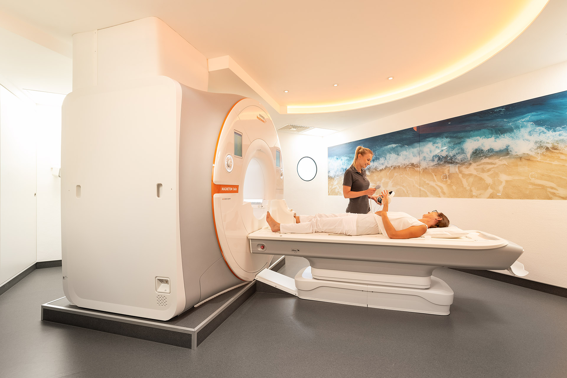
Semi-open MRI machines
If possible, we examine patients with claustrophobia using MRI machines with a particularly large diameter of the "tube", which are therefore referred to as semi-open magnetic resonance tomographs. These devices belong to the latest generation of MRIs with a particularly high level of patient comfort. At up to 70 centimetres, the tube diameter of our MRTs is significantly larger than that of older devices.
Areas of application of magnetic resonance imaging (MRI)
In our practice, magnetic resonance imaging (MRI) is used to diagnose changes and diseases in the following organs and tissues:
- head
- brain and spinal cord spine
- all joints muscles
- ligaments and cartilage
- arms and legs
- throat soft tissue
- abdominal and pelvic organs
- kidneys and urinary tract
- female pelvic organs
- heart and heart vessels
Special MRI examinations
Greater certainty in breast diagnostics thanks to breast MRI
Recent studies recommend regular, supplementary MRI in high-risk patients. Particularly in dense gland tissue, suspicious changes can often be detected more clearly and thus more reliably with MRI. Breast MRI also plays an important role in the assessment of breast implants as well as surrounding gland tissue – this is possible with conventional mammography only to a limited extent.
MRI plays increasingly important role not only in clarification but also in prevention and screening. Recent studies have shown that, for some tumour types, breast MRI can find up to 50% more suspicious lesions than mammography and ultrasound. Therefore the expert association, American Cancer Society, recommends regular performance of breast MRI in high-risk patients (in addition to mammography if necessary) in its current guidelines for breast cancer. For further information about breast diagnostics, please visit the Centre of Excellence for Breast Diagnostics page.
Prostate MRI is the currently best imaging method for depicting the prostate in order to rule out or detect a tumour. MRI provides exact information about the location and spread of a tumour in a non-invasive procedure.
For further information about prostate diagnostics, screening and prostate cancer diagnostics, please visit the Centre for Prostate Diagnostics page.
Whole-body MRI (WB MRI) enables comprehensive imaging analysis of the entire body during one single examination procedure without radiation exposure.
For further information about whole-body MRI, please visit our screening examinations page.
We are set up to examine the heart using magnetic resonance imaging (MRI) and are certified by the German Radiological Society (DRG) as a centre for cardiovascular imaging. The cardiac MRI is performed in the radiology department at the ISAR Klinikum.
Determining the iron and fat content of the liver is an important component in the internal medical clarification of liver diseases and for the early detection of liver damage. Especially in the case of acquired or congenital iron overload in the liver (hemochromatosis), determining the iron content is essential for further therapy and prophylaxis. Determining the fat content can provide very important information for the early assessment of liver remodeling processes.
Modern MRI procedures now allow a simple, non-invasive determination of the iron and fat content of the liver. With the so-called DIXON method, the radiologist and treating doctor are automatically provided with fast and reproducible quantitative graphics on the iron and fat content. In this way, liver biopsies can be avoided in most cases. This state-of-the-art imaging procedure is offered on our latest-generation Siemens MR device Sola at our Giesing site.
Nowadays MR angiography is the method of choice for depicting arterial vessels. Vessels in the neck and head region as well as vessels of the remaining body can be excellently examined with MRI angiography. The very high spatial resolution of modern machines enables very good and timely detection of changes even in tiny vessels. Thanks to this technical progress, non-invasive MR angiography has taken the place of formerly necessary, risky catheter examinations in diagnostics. The only step necessary for the performance of MRI is the injection of a contrast agent into the patient’s vein, which spreads in the vessels, thus enabling vascular imaging. Examination of a body region (e.g. legs) only takes a few minutes and provides reliable results.
Specialist MRI examinations in the field of musculoskeletal imaging
In direct arthrography, MRI or sometimes also CT examination of a joint is performed after a contrast agent has been injected into the joint. With the aid of this technology, which is indicated for special issues, primarily structures of the joint capsule and the cartilaginous parts of a joint can be assessed more accurately than in conventional MRI. This technology is used most frequently for the hip joint, the shoulder, and the wrist, sometimes also on the ankle.
The contrast agent is injected into the joint with a thin needle, about as big as one used for blood sampling, under sterile conditions and after local anaesthesia under X-ray control. Afterwards, the actual imaging is performed in the MRI machine.
The following risks are possible but very rare results of joint puncture: infection, bleeding or an allergic reaction to the contrast agent or the local anaesthetic.
Our Heimeranplatz location is equipped with a small, very powerful MRI scanner (joint scanner) for examinations of the hand/wrist and foot. During the examination, the patient sits comfortably next to the machine and only their hand or foot is inside the tunnel of the machine.
Frequent questions about the MRI examination
A magnetic resonance imaging examination takes approximately 15 minutes depending on the examination region and issue at hand. It is important that the patient remains quietly lying down during this period. Our friendly medical technical staff is in contact with the patient and monitors him/her at all times during the examination.
During the examination, loud yet normal and harmless banging noises occur due to quickly-alternating magnetic fields. For this reason patients wear hearing protection during the examination.
Intravenous administration of a gadolinium contrast agent is required in some cases. It enables more accurate diagnoses and conclusive examination results. Side effects of contrast administration such as an allergic reaction are very rare. For the administration of MRI contrast agents, we use only “macro-cyclical” contrast agents belonging to the group of the safest and most well tolerated MRI contrast agents at all our locations, except for special liver examinations.
Patients with medical devices such as heart pacemakers, brain pacemakers, older heart valves or certain pain pumps usually cannot be examined by MRI since risks for the patients may occur. If you have a medical device and do not know if you can have an MRI examination, please contact us. We will try to clarify this together with you.
Things to know before taking an MRI
If you carry/wear a medical device, please bring the device licence with you to the examination, even if you have already had an MRI examination before. Especially if you have a heart pacemaker or an implanted defibrillator, we ask you to please mention this when requesting an appointment over the phone. We usually carry out an individual check to determine if an MRI examination can be performed.
Further, MRI examinations should not be performed during the first 3 months of pregnancy.
We will be happy to advise you.
Do you have a heart pacemaker or other implanted device and do not know if you can have an MRI examination or have other questions about magnetic resonance imaging?
We are more than happy to answer your questions so please do not hesitate to give us a call via: +49 (0)89 . 550 596 0 or write to us using the contact form.
Appointment and Contact
Our reception team will be glad to help you with all organisational questions. We are further happy to answer your medical questions – before and after the examination.
Get in touch with us: by phone, by making an appointment online or if you have any questions via our contact form.
*For our online appointments we are using a service of the company Doctolib GmbH, Berlin.